
 Dr. Rong Han, Managing Editor, Scientific Editing and Translation
 August 2021
Image manipulation is the process of altering a picture-format image to a desired appearance. In scientific publishing, as almost all journals now require figures to be submitted digitally, image manipulation is typically done using a computer program.
Modifying an image is often necessary for a researcher to generate publication-quality figures, but inappropriate manipulation of images can lead to paper rejection and doubts regarding research credibility. Even though intentional fraud is relatively rare, inappropriate manipulation of images due to ignorance is often seen. The Journal of Cellular Biology (JCB) was among the first journals to rigorously examine images and screen for image manipulation. The guidelines1 developed by The Rockefeller University Press, the publisher of the JCB, have been widely used since its publication. Other journals and organizations have also provided their guidelines and instructions.2-5
Below we summarize the major points of what is allowed and what should be avoided when you prepare your images for publication.
Acceptable image manipulation
The following basic adjustments are about the only manipulations you are allowed to complete to make a picture look better for your manuscript. More complicated processing must be described in the paper itself.
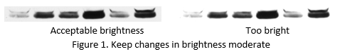
1. Moderate, linear changes applied to the entire image. Linear changes mean brightness, contrast, and color balance revisions. Any change should be applied to the whole image, not a local area. All changes should also be moderate so that they do not cause changes in the interpretation of the results. For example, if you increase the brightness too much, you might make too many pixels have the maximum brightness and therefore lose the subtle differences among these pixels (Fig. 1). Avoid using an image program’s auto contrast, auto levels, and auto color tools, as these may overprocess your image.
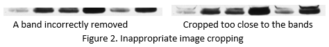
2. Cropping. We often do not need to show the entire picture we receive from the camera or scanner, so it is generally allowed to crop an image to remove unnecessary information or empty space around the edges. However, cropping should not alter the results or introduce ambiguity. Let’s again use the western blot in Fig. 1 as an example. While it is customary to show only the protein band of interest, removing an unwanted band by cropping is not correct. Cropping too close to the band may make readers suspect that you are trying to cover up the fact that there is another band nearby (Fig. 2). Some journals require that a certain area of the blot above and below the band must be shown in the figure.
3. Re-sizing. An image published in a journal article is almost never the same size as the original picture. Re-sizing is a necessary step to ensure your figure meets the journal-desired size. It is generally acceptable to make an image smaller (i.e., decreasing the number of pixels). Computer software can combine multiple pixels into one pixel by averaging their values, and the human eye will not detect a difference. However, do not try to increase the number of pixels of an image, because the computer software will have to create extra pixels that are not already there. This causes misinterpretation of your data and does not make a low-resolution image look better.
Before re-sizing, a scale bar should be added to the image so that you have a correctly sized scale bar for the final figure. A group of images to be shown together for comparison should be re-sized the same way.
1. Plan ahead. If you have planned your experiments well, you are less likely to find it necessary to rearrange or manipulate your figure(s) for publication.
2. Take the best pictures. Get familiar with the instrument and software you use for image acquisition, so that the pictures you take are close to the way they should appear in your paper. Take pictures at the highest resolution possible and save in the correct format (TIFF, not JPEG). You should also save files in the native file format of the image acquisition software because such files may contain metadata of instrument settings such as the magnification information.
2. Take the best pictures. Get familiar with the instrument and software you use for image acquisition, so that the pictures you take are close to the way they should appear in your paper. Take pictures at the highest resolution possible and save in the correct format (TIFF, not JPEG). You should also save files in the native file format of the image acquisition software because such files may contain metadata of instrument settings such as the magnification information.
Also remember…
Always save the original image files. Save another copy when you start processing the image so that you always have the original file. One, if you overprocess or make some other mistakes, you can always start over. Two, journal editors may ask to see your original images to make sure that images have been properly handled. Three, a digital image contains information beyond what we can see with our eyes. An 8-bit grayscale image can have 256 shades of gray, while humans are only able to distinguish about 30 shades of gray. The adjustments we do aim for the best visual effects for human readers, and as a result, the image data are altered, and some information may be lost. If you need to do quantitative measurements, you should use the original images, not the processed ones.
References
1. Rossner M, Yamada K. What’s in a picture: the temptation of image manipulation. J Cell Biol. 2004;166:11-15. Doi:10.1083/jcb.200406019
2. Nature. Image integrity and standards. https://www.nature.com/nature-portfolio/editorial-policies/image-integrity (Accessed August 5, 2021).
3. Office of Research Integrity. Guidelines for best practices in image processing. https://ori.hhs.gov/education/products/RIandImages/guidelines/list.html (Accessed August 5, 2021)
4. Cromey DW. Avoiding twisted pixels: ethical guidelines for the appropriate use and manipulation of scientific digital images. Science and Engineering Ethics. 2010;16:639-667. https://doi.org/10.1007/s11948-010-9201-y
5. Sakabe K. American Society for Biochemistry and Molecular Biology series Due Diligence. ASBMB Today. https://www.asbmb.org/asbmb-today/authors/kaoru-sakabe (Accessed August 5, 2021) or https://www.asbmb.org/getmedia/2af208df-ae20-4da0-b347-0180de6238bb/due-diligence.pdf